 Autoantibodies & Altered Antiviral Immune Responses in a Subset of Children With PANS
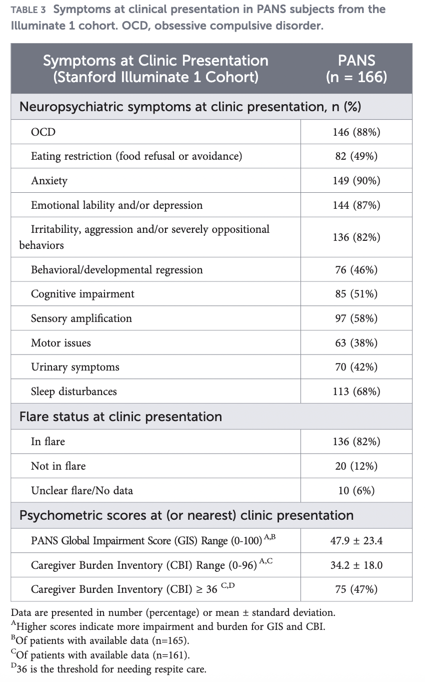
A recent study conducted by researchers at Stanford University offers further evidence of immune dysregulation and autoimmunity in Pediatric Acute-onset Neuropsychiatric Syndrome (PANS). The investigators,
-
Identified increased prevalence of autoantibodies linked to established autoimmune diseases in patients with PANS.
-
In a smaller subset of patients, they discovered autoantibodies that can interfere with a critical component of the body’s antiviral immune defense.
Published in Frontiers in Immunology in July 2026, the study analyzed 224 plasma samples collected during PANS flares from 166 patients and compared them with samples from 83 healthy pediatric controls. The findings do not establish a new diagnostic test for PANS or demonstrate causality between these autoantibodies and PANS. Rather, the results contribute to a growing body of research indicating that immune abnormalities are present in PANS and suggest that the underlying immune mechanisms may vary among patients.
Evidence of Broader Immune Dysregulation
Researchers examined antibodies against 27 targets associated with established autoimmune diseases, including systemic lupus erythematosus, Sjögren syndrome, dermatomyositis, scleroderma, autoimmune thyroid disease, pernicious anemia, and autoimmune vasculitis.
Children with PANS were significantly more likely than healthy controls to possess certain autoantibodies associated with scleroderma and gastrointestinal or endocrine autoimmune diseases. The PANS cohort also had a high rate of clinical and laboratory findings associated with immune dysregulation.
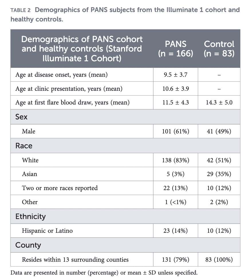
Of the 166 patients,
-
30% exhibited arthritis
-
36% presented with livedo reticularis (a skin finding that causes a net-like or lace-like pattern of reddish-blue or purple discoloration)
-
18% exhibited periungual redness (redness around the nail folds). The authors note that periungual redness and swelling are classic findings associated with scleroderma and dermatomyositis and were among the clinical signs of autoimmunity evaluated in the PANS cohort.
Among those who underwent specific laboratory testing, 33% had low complement C4, 22% had a positive ANA at a titer of at least 1:80, and 19% had elevated antithyroid antibodies.
Overall, 65% of patients with available data exhibited an autoimmune or inflammatory disease or finding beyond arthritis. Diagnosed autoimmune and inflammatory conditions within the cohort included thyroiditis, psoriasis, celiac disease, inflammatory bowel disease, chronic urticaria, eosinophilic esophagitis, lupus, and additional disorders. Patients with connective-tissue-disease-associated autoantibodies identified by the researchers were nearly three times more likely to have another autoimmune condition after adjustment for age, sex, and race. However, the presence of these autoantibodies was not associated with flare duration, arthritis, or signs of vasculopathy. The significant clinical association was with the presence of other autoimmune conditions.
These results do not indicate that children with PANS have scleroderma or another specific autoimmune disease solely due to the presence of these antibodies. Instead, the observed pattern may reflect a broader predisposition toward immune dysregulation or loss of normal immune tolerance in some patients with PANS.
Understanding Autoantibodies
Normally,
-
antibodies recognize viruses, bacteria, and other substances the immune system identifies as foreign.
-
Autoantibodies mistakenly recognize something belonging to the person’s own body.
Autoantibodies are present in many established autoimmune diseases; however, their detection does not necessarily indicate causation. Some autoantibodies may primarily serve as markers of abnormal immune activity, while others can directly disrupt normal biological functions. The researchers therefore extended their investigation beyond antibody identification to assess whether certain autoantibodies could interfere with the function of their target proteins.
Autoantibodies Targeting IFN-λ
Researchers identified a subset of children with antibodies against interferon lambda (IFN-λ). Interferons are signaling proteins used by the immune system to coordinate responses to infection. IFN-λ, also called type III interferon, is particularly important at the body’s barrier surfaces, including the respiratory and gastrointestinal tracts, skin, lungs, and blood-brain barrier.
When a virus encounters these surfaces, IFN-λ assists surrounding cells in establishing an antiviral state, thereby impeding viral spread. The identification of IFN-λ autoantibodies is therefore potentially significant; however, the mere presence of an antibody does not confirm interference with immune function. Consequently, researchers conducted additional laboratory experiments to determine whether the antibodies simply bound to IFN-λ or actively inhibited its function.
Among 11 tested PANS samples with IFN-λ-binding autoantibodies,
-
9 showed detectable inhibition of IFN-λ signaling
-
Four met the researchers’ stricter predefined threshold for IFN-λ-neutralizing activity
-
None of the tested healthy-control samples met that neutralization threshold.
Researchers also identified some patients with antibodies that bound several type I interferons, including IFN-α7, IFN-α8, IFN-α10, and IFN-β. However, functional testing did not demonstrate neutralization of the tested type I interferons, suggesting that the neutralizing activity observed in these experiments was selective for the type III IFN-λ pathway.
The researchers subsequently isolated IgG from samples with high neutralizing activity and tested the components independently. The IgG antibody fraction retained neutralizing activity, whereas plasma depleted of IgG did not, indicating that the antibodies themselves were responsible for blocking IFN-λ signaling in these samples. These experiments demonstrated that, in a subset of samples, the antibodies themselves had a measurable functional effect on an immune pathway.
Why IFN-λ Could Matter in PANS
IFN-λ is especially relevant to PANS because infections are frequently associated with symptom onset and subsequent flares. Unlike other interferons that function systemically, receptors for IFN-λ are primarily concentrated at barrier tissues. Research conducted outside the context of PANS has demonstrated that loss of normal IFN-λ activity can increase susceptibility to infection at these sites.
The authors also highlight another potentially important barrier: the blood-brain barrier. The blood-brain barrier regulates movement of cells and substances from the circulation into the central nervous system, and experimental research has demonstrated that IFN-λ can help strengthen this barrier during viral infection. In a mouse model of West Nile virus infection, for example, IFN-λ signaling tightened the blood-brain barrier and helped limit viral entry into the central nervous system.
Neuroinflammation and disruption of blood-brain barrier integrity have been proposed as potential components of the PANS disease process. Previous neuroimaging and related studies have identified abnormalities in brain regions such as the basal ganglia. The findings from this study therefore raise the question of whether impaired IFN-λ signaling in some patients could compromise the body’s ability to control infection or inflammation at barrier tissues, including the blood-brain barrier.
The current study does not establish a direct connection, nor does it demonstrate that IFN-λ autoantibodies cause blood-brain barrier disruption in PANS. The findings instead identify a potential biological pathway linking infection, immune dysfunction, and neuroinflammation that warrants further investigation.
Another Potential Target: The IFN-λ Receptor
Researchers also looked for antibodies against IFNLR1, part of the receptor that cells use to respond to IFN-λ. Several PANS patients had unusually high antibody binding to this receptor, and in one patient, researchers demonstrated that the antibodies could interfere with IFN-λ signaling through the receptor.
The authors note that IFNLR1-neutralizing autoantibodies have not been previously described. As functional neutralization was identified in only one patient, no association between these antibodies and PANS can currently be established. The finding warrants further investigation of IFNLR1 as another potential target within this immune pathway.
Autoantibody Levels Did Not Track With PANS Flares
Researchers also examined samples collected from some patients at different points in time and found that IFN-λ autoantibody levels did not consistently rise during PANS flares or fall during recovery. Some patients had their highest levels during flares, while others had higher levels during periods of improvement or recovery.
This finding suggests that measuring circulating IFN-λ-binding antibodies alone is unlikely to serve as a reliable indicator of current PANS flare status. Importantly, antibody binding and biological function were not equivalent. Individuals may possess antibodies that bind the same immune target without those antibodies exerting identical effects. In this study, the degree of IFN-λ antibody binding did not consistently predict the extent to which patient plasma inhibited IFN-λ signaling, indicating that future research should assess both antibody levels and their functional consequences.
What the Study Does and Does Not Tell Us
The findings are significant; however, they require cautious interpretation. This study does not demonstrate that IFN-λ autoantibodies cause PANS, nor does it establish that these antibodies are prevalent among most individuals with PANS.
What the researchers found
-
Functionally active IFN-λ-neutralizing autoantibodies have been identified in a subset of patients with PANS.
-
Laboratory analyses demonstrated that some of these antibodies can interfere with IFN-λ signaling, which is a critical component of the antiviral immune response.
-
These findings support further investigation into whether impaired antiviral defenses contribute to disease mechanisms in a subset of patients.
What the results do not mean
No statistically significant difference was observed between the PANS and healthy control groups regarding the overall prevalence of antibodies that bind to the IFN-λ pathway.
Functional testing was conducted on a small, intentionally selected group of samples exhibiting higher antibody binding. Among the 11 PANS samples tested, nine demonstrated some inhibition of IFN-λ signaling, and four met the study’s more stringent criteria for neutralization. None of the 11 selected healthy-control samples met that threshold. However, the difference between the four PANS samples and zero healthy controls did not itself reach statistical significance (p=.09). These results therefore cannot be extrapolated to estimate the prevalence of IFN-λ-neutralizing autoantibodies in the broader PANS population and require confirmation in larger cohorts.
The study does not establish a causal relationship between these antibodies and PANS, nor does it explain PANS symptoms. Children with IFN-λ autoantibodies could not be clinically distinguished from other children with PANS.
The PANS and healthy-control groups also differed in age and racial composition. The authors considered these demographic differences when interpreting the results, but limited subgroup sizes prevented robust adjustment for these factors across all individual autoantibody analyses, IFN-λ-neutralizing autoantibodies, and detailed clinical phenotypes.
Not a diagnostic test
The autoantibodies identified in this study should not be regarded as diagnostic biomarkers for PANS at this time. Their significance for individual patients remains unclear. Some may be associated with existing autoimmune conditions, others could precede the development of additional autoimmune diseases, and some may indicate broader immune dysregulation.
Larger and longer-term studies are required to determine the prevalence of these functionally active antibodies in PANS, to identify which patients develop them, and to assess whether they contribute to disease mechanisms or clinical outcomes.
Where the Research Goes From Here
This study adds to evidence of immune dysfunction and autoimmunity in PANS while providing further insight into the specific immune pathways involved and their potential functional consequences.
PANS is a clinically heterogeneous condition. Patients may present with diverse triggers, symptoms, disease courses, immune findings, and responses to treatment. It is increasingly likely that PANS will not be explained by a single immune abnormality or disease mechanism. Instead, research may identify biologically distinct subgroups within the broader PANS population, with different immune pathways contributing to illness in different patients.
The authors call for larger studies combining detailed clinical information with immune, genetic, and protein profiling to investigate these potential subgroups. They also recommend studying IFN-λ-neutralizing autoantibodies in paired blood and cerebrospinal fluid samples, which could help determine whether these antibodies are present within the central nervous system and whether they have any relationship to neuroinflammation. Future research can also investigate whether disruption of IFN-λ signaling affects blood-brain barrier function and susceptibility to infection or inflammation.
For patients and families, this research does not provide a new test or an immediate change in treatment. What it provides is additional evidence that immune abnormalities observed in PANS are not limited to nonspecific laboratory findings. Researchers identified an increased prevalence of autoantibodies associated with established autoimmune diseases, substantial autoimmune and inflammatory findings within the study population, and, in a subset of patients, antibodies with the demonstrated ability to interfere with an antiviral immune pathway.
Studies like this are beginning to examine how measurable immune abnormalities may actually function biologically and how abnormalities involving infection response, autoimmunity, barrier immunity, and neuroinflammation could intersect in PANS. While much remains to be established, identifying specific, functionally altered immune pathways provides researchers with concrete mechanisms to investigate and represents an important direction for PANS research.
Study: Yin X, Frankovich J, Kalaycioglu M, et al. Interferon-λ-neutralizing autoantibodies and common autoimmune disease autoantibodies in pediatric acute-onset neuropsychiatric syndrome. Frontiers in Immunology. Published July 17, 2026.
https://www.frontiersin.org/journals/immunology/articles/10.3389/fimmu.2026.1832833/full
Gabriella True
